Animal Cell Through Light Microscope / Magnification Bioninja - Human cheek epithelial cells stained with methylene blue @400xtm.. All the living matter of a cell is called protoplasm. Plant cells have cell walls, one large vacuole per cell, and chloroplasts, while animal cells will have a cell membrane only. These structures are discussed in more detail in the following pages. .cell with an animal cell, as seen under a light microscope, limited to cell wall, nucleus, cytoplasm, chloroplasts, vacuoles and location of the cell membrane. Human cheek epithelial cells stained with methylene blue @400xtm.
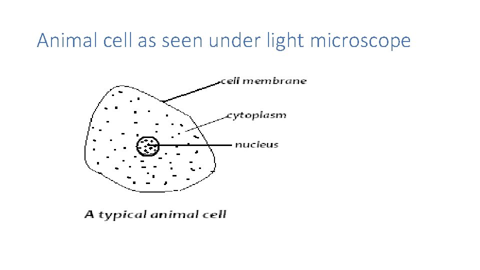
The diagram below is an animal as may be seen using a light microscope. 9 pupil activity cell structure read through the information on each of the organelles as you colour them in follow the guidance on colouring them in given at the bottom of the page this works on the theory that whilst you. All the living matter of a cell is called protoplasm. Plant cells have cell walls, one large vacuole per cell, and chloroplasts, while animal cells will have a cell membrane only. Plant cells, animal cells and bacteria can be visualized through the light microscope.
Components Teacher Stem In Curriculum Mr Mwaurah School from slidetodoc.com With a light microscope you can see several structures inside the cell. An animation that shows animal cells. A cell is a very tiny structure which exists in living bodies. In order to view microorganisms that do not produce much pigment, you must use phase contrast or gram stain (methylene blue is another option). Cheek cells under a microscope. Mixed slide of stained protozoa 100xtm. Light microscopes use a number of lenses to produce an image that can be viewed directly at the eyepiece. Cells in animals and plants.
It is the simplest type of microscope.
It is the simplest type of microscope. Animal cell through a microscope. (reproduced by permission of photo. The substage condenser directs light through the slide into the objective. The top lens through which you look is called the eyepiece while the lower lens that is close to the slide is called the objective lens. All the living matter of a cell is called protoplasm. You can view leaf cells using the microscope. Alternatively, candidate cells for imaging can be located using brightfield, phase contrast. Always begin by viewing the object through a low power lens. Light microscope uses the properties of light to produce an enlarged image. Plant cells, animal cells and bacteria can be visualized through the light microscope. With a light microscope you can see several structures inside the cell. This is an example of what you might see when you look at plant cells through a light microscope.
Their structure and composition, and how they work. Cell membrane, cytoplasm and nucleus that can observed through light microscope. With a light microscope you can see several structures inside the cell. Mdcat biology live lecture 1, ch no 1, light and electron microscope + animal and plant cells. The top lens through which you look is called the eyepiece while the lower lens that is close to the slide is called the objective lens.
71 744 Plant Cell Stock Photos Pictures Royalty Free Images Istock from media.istockphoto.com Slides and a microscope is an important instrument for studying cells e.g. Albeit the detail will be minimal without a you are observing two unlabeled cells, a plant and an animal cell, through a microscope. Human cheek epithelial cells stained with methylene blue @400xtm. They are called compound microscopes. You can view leaf cells using the microscope. Animal cell features (light microscope). Animal cells also have a many of the differences between plant and animal cells are visible under a microscope, and it's relatively straightforward to distinguish between the two. People's surprise, mitochondria are visible in the light microscope.
An animation that shows animal cells.
Plant cell (onion cell) and animal cell (cheek cell) can be observed under a light microscope. .cell with an animal cell, as seen under a light microscope, limited to cell wall, nucleus, cytoplasm, chloroplasts, vacuoles and location of the cell membrane. (reproduced by permission of photo. People's surprise, mitochondria are visible in the light microscope. The top lens through which you look is called the eyepiece while the lower lens that is close to the slide is called the objective lens. This is an example of what you might see when you look at plant cells through a light microscope. These structures are discussed in more detail in the following pages. Light microscopy (the use of microscopes is called microscopy) Human cheek epithelial cells stained with methylene blue @400xtm. Cell membrane, cytoplasm and nucleus that can observed through light microscope. Plant cells have cell walls, one large vacuole per cell, and chloroplasts, while animal cells will have a cell membrane only. Light microscopes are used in biology classes in schools and colleges as well as in professional scientific environments such as government laboratories and biotechnology companies. 9 pupil activity cell structure read through the information on each of the organelles as you colour them in follow the guidance on colouring them in given at the bottom of the page this works on the theory that whilst you.
Human cheek epithelial cells stained with methylene blue @400xtm. They are called compound microscopes. Organisms are made up of cells. Light microscopes use a number of lenses to produce an image that can be viewed directly at the eyepiece. Light passes from a bulb under the stage, through a condenser lens and then through when you look at animal or plant cells under the electron microscope, you can see a lot more detail.
Difference Between Plant And Animal Cells Cells As The Basic Units Of Life Siyavula from intl.siyavula.com Cheek cell and oral bacteria @100xtm. With light microscopy i can simply scrape some cells from my cheek smear them on a slide and look at them. You can view leaf cells using the microscope. A cell is a very tiny structure which exists in living bodies. Cheek cells are eukaryotic cells (cells that contain a nucleus and other organelles within enclosed in a although the entire cell appears light blue in color, the nucleus at the central part of the cell is much darker, which. These structures are discussed in more detail in the following pages. Image:animal cell seen under light microscope. People's surprise, mitochondria are visible in the light microscope.
Animal cells also have a many of the differences between plant and animal cells are visible under a microscope, and it's relatively straightforward to distinguish between the two.
All living things are composed of cells. Light microscope uses the properties of light to produce an enlarged image. 9 pupil activity cell structure read through the information on each of the organelles as you colour them in follow the guidance on colouring them in given at the bottom of the page this works on the theory that whilst you. Light microscopes use a number of lenses to produce an image that can be viewed directly at the eyepiece. Plant cells, animal cells and bacteria can be visualized through the light microscope. The diagram below is an animal as may be seen using a light microscope. Cell membrane, cytoplasm and nucleus that can observed through light microscope. A cell is a very tiny structure which exists in living bodies. Trypanosoma, cause of african sleeping sickness @400x tm. We started this unit with an overview of cells: Observing the cells through the microscope eyepieces takes several seconds, which is at least tenfold longer than is often required to obtain an image of sufficient quality for cell selection and focusing. Three main parts can be seen: You can view leaf cells using the microscope.
Share :
Post a Comment
for "Animal Cell Through Light Microscope / Magnification Bioninja - Human cheek epithelial cells stained with methylene blue @400xtm."


Post a Comment for "Animal Cell Through Light Microscope / Magnification Bioninja - Human cheek epithelial cells stained with methylene blue @400xtm."